Anti Lamin Immunofluorescence

Lamin B1 Antibody 119d5 F1 Nuclear Envelope Marker Ab8982 Abcam

Alexa Fluor 647 Anti Lamin B1 Antibody Epr8985 B Ab194108 Abcam

Alexa Fluor 488 Anti Lamin B2 Antibody Epr9701 B Ab200426 Abcam

Anti Lamin A Antibody Ko Tested Ab26300 Abcam

Alexa Fluor 594 Anti Lamin A Lamin C Antibody Epr4100 Ab215324 Abcam

Alexa Fluor 647 Anti Lamin B2 Antibody Epr9701 B Ab200427 Abcam

Anti lamin a c terminal antibody is suitable for use in western blot 0 1 0 2 μg ml using hela nuclear extract.
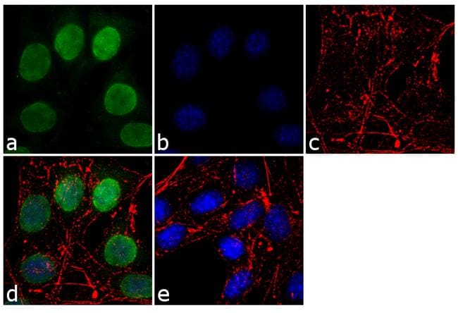
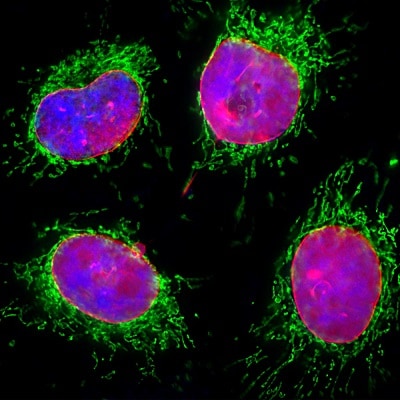
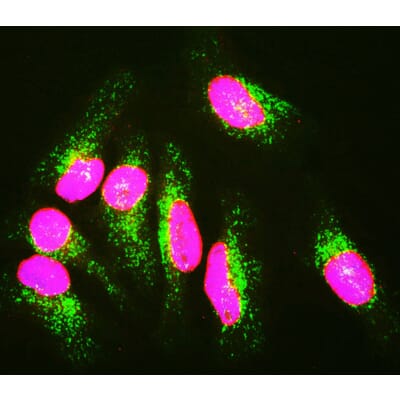
Anti lamin immunofluorescence.

7497 Phospho Ndrg1 Thr346 D98g11 Xp Rabbit Mab Alexa Fluor 647 Conjugate Cst抗体 Confocal Immunofluorescent Analysis Of Mab Painting Alexa

Alexa Fluor 488 Anti Lamin A Lamin C Antibody Ep4520 16 Ab205769 Abcam

Recombinant Anti Lamin B1 Antibody Epr22165 121 Bsa And Azide Free Ko Tested Ab239399

Recombinant Anti Lamin B1 Antibody Epr8985 B Ko Tested Ab133741 Abcam

Anti Lamin A Lamin C Antibody Wl4g10 Ko Tested Ab232730 Abcam

Alexa Fluor 488 Anti Lamin A Lamin C Antibody Epr4100 Ab185014 Abcam
Lamin B1 Antibody Pa5 19468

Anti Lamin A Lamin C Antibody Wl4g10 Bsa And Azide Free Ko Tested Ab269575
Anti Lamin A Lamin C Antibody 4c4 Ko Tested Ab190380 Abcam

Alexa Fluor 647 Anti Lamin B Receptor Lbr Antibody E398l Ab201349 Abcam

Anti Laminin 1 2 Antibody Ab7463 Abcam

Anti Lamin A Lamin C Antibody 4c11 Bsa And Azide Free Ko Tested Ab244577

Anti Lamin B1 Antibody Nuclear Envelope Marker Ko Tested Ab16048 Abcam

Cell Signaling Anti Phospho Dakt 1 200

Alexa Fluor 647 Goat Anti Mouse Igg Secondary Antibody Ab150115 Abcam

Anti Lamin B2 Antibody Ln43 Ab8983 Abcam

Anti Pcna Mouse Monoclonal Antibody 1d7 Abfluor 555 Conjugated Life Science Eukaryotic Cell Cell Cycle

Abcam Anti Erk1 Erk2 Phospho T185 Y187 T202 Y204 Mapk Yt Anti Microscopy Human

Recombinant Anti Lamin B1 Antibody Epr8985 B Bsa And Azide Free Ko Tested Ab220797

Lamin A Atto 647n Antibody Immunofluorescence L3544 Anti Lama Sigma Aldrich
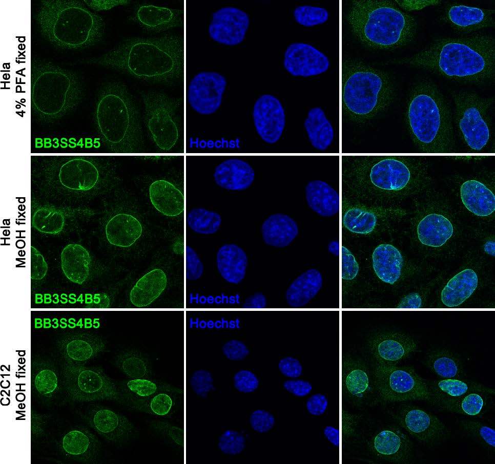
Anti Lamin B Receptor Bb3ss4b5 Monoclonal Antibodies Ximbio

Anti Lamin B Receptor Lbr Antibody Bbmlbr 12 F8 Ko Tested Ab232731 Abcam

Alexa Fluor 488 Anti Lamin B Receptor Lbr Antibody E398l Ab201532 Abcam
Anti Lamin A C Antibody A85443 Antibodies Com
Source : pinterest.com
